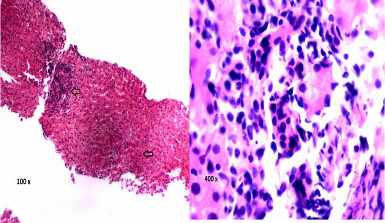
Figure 1.
Liver histology on H&E staining (×100) from a percutaneous liver biopsy obtained on day 8 of hospitalisation in a patient with unexplained hepatitis. The arrow shows an area of periportal and lobular inflammation. At ×400 magnification, the area of periportal inflammation can be identified being comprised of mononuclear cells. No cytomegalovirus inclusion bodies were identified.

