
Figure 2.
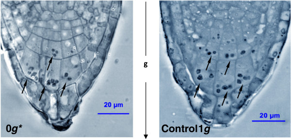
The root columella cells observed by phase-contrast microscopy. (Left-hand image) Samples were fixed, embedded in resin and 2 μm semithin sections were obtained from them and observed under phase-contrast light microscopy. Those samples grown under magnetic levitation (0 g* tube) for 4 days show the same distribution of statoliths (indicated by arrows) as in control 1 g samples (right-hand image) grown outside the magnet. The statoliths collect near the basal membrane of the columella cells, under the force of gravity; the arrow between the images indicates the direction of gravity.