Figure 1.
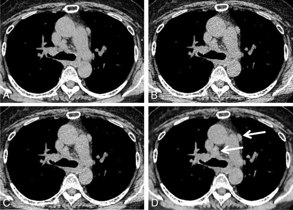
Transverse chest CT through the ascending aorta in a 64 year-old woman with mediastinal lymph node enlargement (arrows). Images were obtained with standard-dose FBP CT (A), low-dose FBP CT (B), low-dose ASIR 50% (C), and low-dose MBIR method (D). Note the excellent depiction of mediastinal lymph nodes on the low-dose MBIR image (lesion conspicuity score 5), compared with low-dose CT with FBP (score 3) and ASIR (score 4). Objective image noise on low-dose MBIR CT is 12.12 HU, showing higher than those on standard-dose FBP (17.72 HU), low-dose FBP (27.69 HU), and low-dose ASIR CT (20.76 HU) in this patient.

