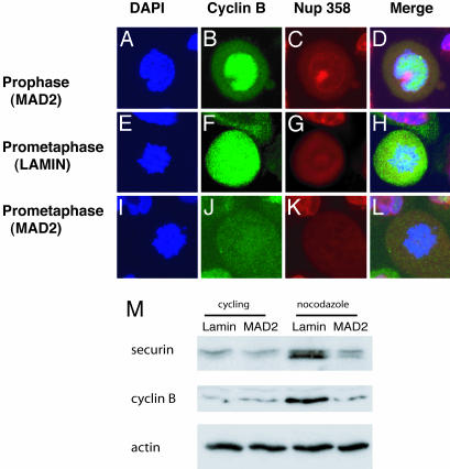
Fig. 4.
Premature cyclin B degradation in MAD2 knockdown HaCaT cells. Immunofluorescence microscopy showing nuclei (blue), cyclin B (green), nuclear envelope-Nup 358 (red), and merged images (×100). (A–D) MAD2 knockdown cells show high levels staining of nuclear cyclin B during prophase, during which the nuclear envelope is still visible similar to lamin controls (data not shown). (E–H) Lamin knockdown control cells stain intensely for cyclin B during prometaphase, and the nuclear envelope is dissolved at this stage of mitosis. (I–L) MAD2 knockdown cells with prometaphase morphology lacking cyclin B. (M) Immunoblot analysis of securin and cyclin B protein levels after 16 h of exposure to nocodazole comparing lamin and MAD2 knockdown HeLa cells.

