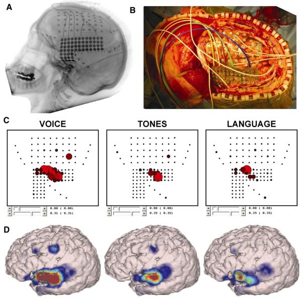
Fig. 4.
Real-time assessment of auditory and receptive language nodes: patient with epilepsy with ECoG electrodes implanted over left frontal, parietal, and temporal cortex. A lateral X-ray (A) and an operative photograph (B) depict the configuration of two grids (one 40-contact frontal grid, one 68-contact temporal grid) and three 4-contact strips. A passive mapping procedure (SIGFRIED) identified eloquent language cortex by contrasting task-related changes during listening to voices and tones (C, D). The results are presented in two intuitive interfaces: a two-dimensional interface that mimics the electrode grid and a three-dimensional anatomically correct interface.

