
Fig. 6.
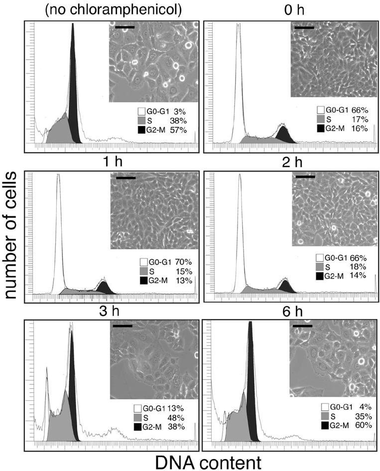
Effect of bacterial protein synthesis inhibition on the display of CDT activity by S. typhi. Cultured intestinal Henle-407 cells were infected with the S. typhi strain, and the bacterial protein synthesis inhibitor chloramphenicol was added at different times after infection as indicated. Cells were examined 72 h after infection under a phase microscope for signs of intoxication or processed to measure DNA content by flow cytometry as indicated in Materials and Methods. The peaks corresponding to cells in G0/G1 (G0-G1), S, or G2 (G2-M) are indicated. (Scale bar, 50 μm.)