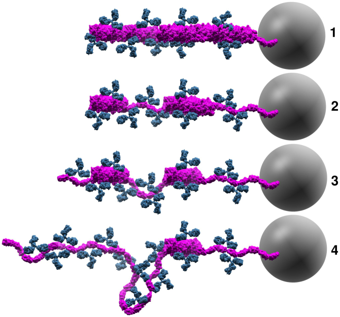
Figure 7. Conceptual model of antibodies attaching to P-fimbriae.
A schematic representation of our conceptual model for molecular events monitored in the force spectroscopy experiments shown in Fig. 3. The panels represent the configuration of the fimbria (magenta) and antibodies (blue) prior to, and during, the extension phase. Antibodies initially interacted with the helical shaft. After the fimbria was unwound some antibodies prevented proper rewinding by interfering with the LL interactions. Loops were able to form when antibodies bound two subunits in an unwound configuration. The gray sphere illustrates the trapped 2.5 μm bead (not drawn to scale) that was used as probe in the force spectroscopy experiments.

