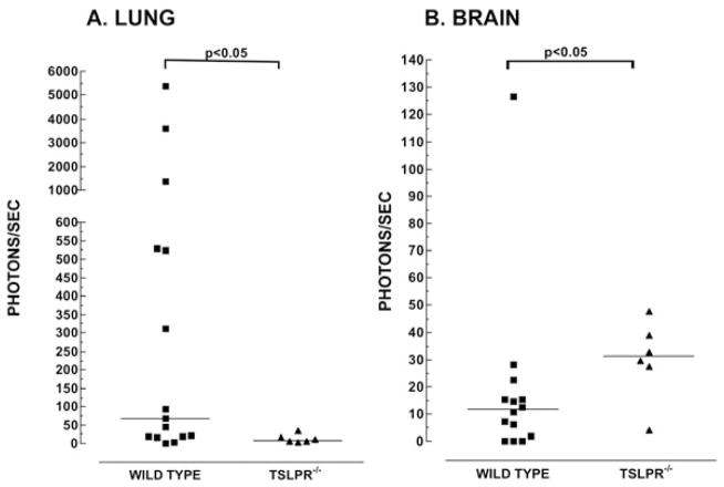
Figure 3. The relative median number of tumor cells is lower in the lung and higher in the brain in Tslpr−/− mice.
Tumor-bearing mice that had reached their humane endpoint were euthanized and the lungs and brains were assayed by chemiluminescence for the presence of 4T1-12B cells. Symbols represent the number of photons/sec measured for a whole organ lysate obtained from an individual WT (■) or Tslpr−/− (▲) mouse. Medians, denoted by a horizontal line, were compared using a Mann-Whitney test. The number of mice in each group were as follows: Lung: n=15 for WT and n=6 for Tslpr−/−. Brain: n= 14 for WT and n=6 for Tslpr−/− mice. Data shown in Figures 6A and 6B are from three separate experiments, combined.

