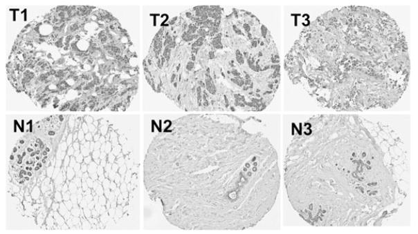
Figure 7. TSLP is expressed in both breast cancer cells and in normal, glandular breast epithelial cells.

Tissue microarrays (TMA) were prepared from formalin-fixed, paraffin-embedded tissue from the Manitoba Breast Tumor Bank. TSLP expression (staining brown) is present in both breast tumors (T) and normal breast tissue (N) that had been procured from mammoplasty reduction surgery. Representative cores from three different patients (T1, T2 and T3) are shown, along with representative cores of normal breast tissue from three individuals (N1, N2 and N3). A total of 50 normal breast tissue cores and 50 malignant breast tissue cores were present in the TMA that was analyzed.
