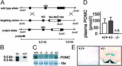
Fig. 1.
Targeted deletion of the murine Pomc locus. (A) Schematic diagrams and partial restriction maps of the mouse wild-type Pomc locus, Pomc targeting vector, and mutant Pomc allele. The filled boxes represent Pomc coding sequence or 5′ probe. The open boxes represent the tau-lacZ-PGK-neor cassette. The loxP sites (not shown) are in direct orientation such that a Cre-mediated recombination event would cause removal of the PGK promoter and neor coding sequence. (B) Southern blot analysis of tail DNA from wild-type and Pomc+/- mice digested with EcoRI. The 9.5-kb band represents the wild-type allele whereas the lower, 6.5-kb band represents the targeted allele. (C) Northern blot analysis showing an absence of POMC mRNA expression in brains from Pomc-/- mice. (Lower) UVL photograph of 18s RNA as loading control. (D) Two-site immunoassay showing that peripheral POMC peptides are not detectable (n.d.) in plasma from Pomc-/- mice. (E) Histochemical staining for β-galactosidase activity in brains from Pomc+/- mice demonstrated POMC-expressing neurons projecting from the arcuate nucleus to hypothalamic and extra-hypothalamic sites. No staining is observed in brains from wild-type mice.

