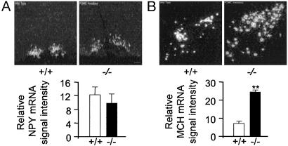
Fig. 4.
Hypothalamic NPY and MCH mRNA expression in Pomc-/- and wild-type mice. (A) Pomc-/- mice have normal levels of NPY mRNA in the arcuate nucleus but an absence of NPY in the DMH. Shown are representative darkfield photomicrographs of the arcuate nucleus from male wild-type and Pomc-/- mice. (Scale bar = 150 μm.) Graphed data represent mean of four consecutive brain sections taken from a single brain. Four brains of each genotype were analyzed. (B) MCH mRNA levels are elevated in the lateral hypothalamus of Pomc-/- mice. Shown are representative darkfield photomicrographs of the lateral hypothalamus in a wild-type and Pomc-/-. (Scale bar = 200 μm.) Graphed data represent mean of six consecutive brain sections taken from a single brain. Five brains of each genotype were analyzed. **, P < 0.01.

