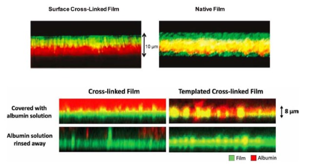
Figure 7.

Laser scanning confocal microscopy cross-sectional images of nanofilm biomaterials formed by the layer-by-layer assembly of charged polymers. Top) A 60-layer red fluorescing film terminated with a green fluorescing activated polymer (i.e., capable of forming chemical cross-links, left) and a green fluorescing standard polymer (right). Green confined to the surface of the film to the left (but not right) suggests cross-links to occur in the surface region. Bottom) Red fluorescing albumin is added to a cross-linked (i.e., non porous) and a templated cross-linked (i.e., porous) film. The albumin penetrates only the porous film, as evidenced by the red and yellow color throughout. Reproduced with permission from The American Chemical Society (top) [24] and John Wiley and Sons (bottom) [25].
