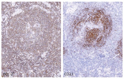
Figure 1.

Nodal marginal zone lymphoma. The panel shows an example of nodal marginal zone lymphoma (MZL) with follicular colonization. BCL-2+ neoplastic cells surround and colonize the germinal center, whereas CD23 highlights the disrupted follicular dendritic cell meshwork. Images were acquired with the Olympus Dot. Slide Virtual microscopy system using an Olympus BX51 microscopy equipped with PLAN APO 2x/0.08 and UPLAN SApo 40x/0.95 objectives.
