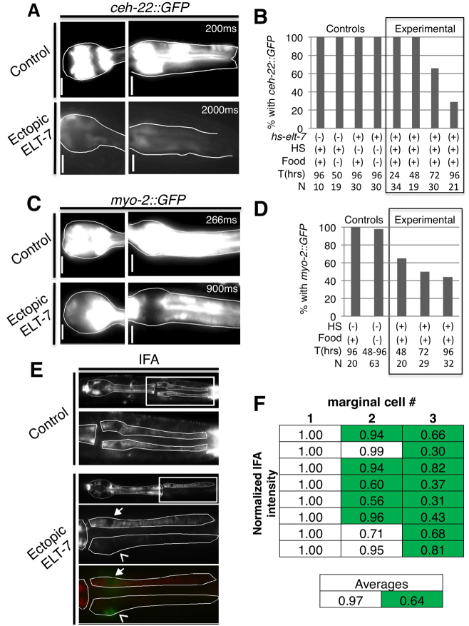
Fig. 3.
Loss of pharynx-specific markers following a pulse of ELT-7 expression. Expression of pharynx muscle-specific ceh-22::GFP (A,B) or myo-2::GFP (C,D) reporters diminishes by 3 days after ectopic ELT-7 expression compared with non-heat shocked hs-elt-7 control larvae. Scale bars: 5 μm. Exposure time is indicated in the upper right-hand corner. (E) Decreased IFA in cells expressing elt-2::lacZ::GFP compared with non-GFP-expressing marginal cells within the same section of the pharynx (boxed region shown in lower panels). Bottom panel, overlay of IFA (red) and elt-2::lacZ::GFP (green). Closed arrow indicates non-elt-2::lacZ::GFP-expressing marginal cell nucleus; open arrow indicates GFP-expressing nucleus. Faint green signal over the non-expressing nucleus is the out-of-plane signal from the positive nucleus of the third marginal cell. (F) IFA average pixel intensity minus background was normalized to the cell with the highest pixel intensity within each worm. Normalized signal in anterior marginal cells with strong elt-2 reporter expression (green highlighted) was lower than in cells with weak or no expression of elt-2 (white). Each row is an individual animal and each column indicates the individual marginal cells ordered based on increasing GFP signal. The average overall IFA signal is reduced in the elt-2::lacZ::GFP-positive marginal cells.

