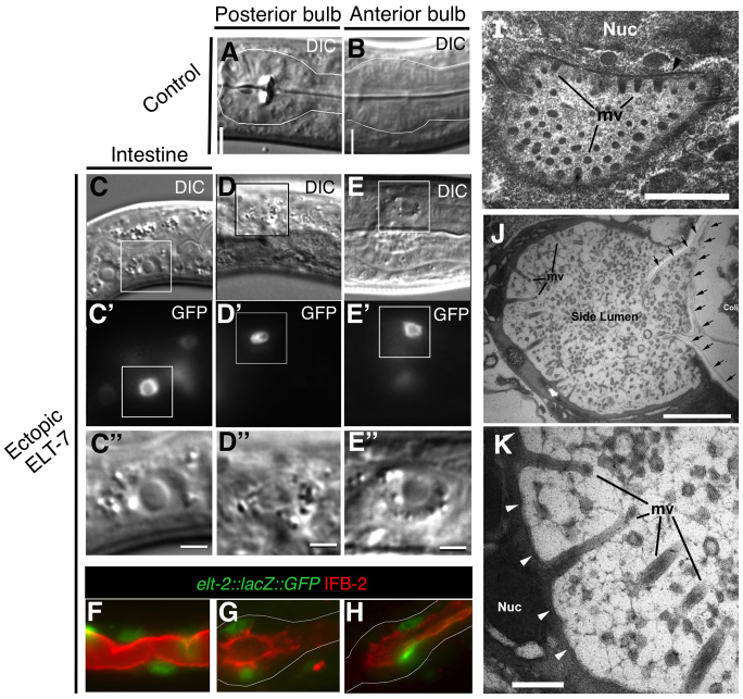
Fig. 4.
Reprogrammed pharyngeal cells have intestine-specific morphology. Morphology of the posterior (A) and anterior (B) bulbs of the pharynx in non-heat shocked hs-elt-7 control animals and morphology of the intestine (C), posterior bulb (D) and anterior bulb (E) after heat shock (D, 72 hours; E, 96 hours) are seen using DIC. Boxed region (C′-E′) outlines elt-2::lacZ::GFP-expressing nuclei in C′-E′. Scale bars: 2 μm. (F-H) Overlay of IFB-2 (red) and elt-2::lacZ::GFP (green) signal in the endogenous intestine (F), and posterior (G) and anterior (H) pharynx 5 days after ectopic ELT-7. (I-K) Morphology of the intestine and remodeled pharynx by TEM. (Extensive images of the ultrastructure of the wild-type pharynx for comparison can be viewed at wormatlas.org.) (I) Cross-section of normal L1 larva intestinal lumen. Microvilli (mv) on the top are seen at nearly full length, whereas those at the bottom are at oblique angles. Black arrow indicates the terminal web. Scale bar: 1 μm. (J) Accessory lumen in the anterior pharynx of hs-elt-7 L1 larva 6 days after brief ectopic ELT-7 expression. Arrows show remaining buccal cuticle. Several long microvilli (mv) are seen and the circular profiles in the middle of the side lumen are apparently additional microvilli extending at extreme angles from other edges of the side lumen. Some E. coli (Coli) lie within the buccal channel. Scale bar: 1 μm. (K) Higher magnification of ectopic microvilli (mv). Scale bar: 0.2 μm. Arrowheads indicate an electron-dense line, an apparent terminal web structure, running subjacent to the plasma membrane. The microvilli appear surrounded by diffuse glycocalyx. Nuc, nucleus.

