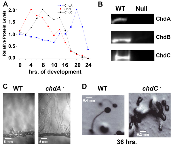
Fig. 1.
The three CHD proteins of Dictyostelium have distinct developmental roles. (A) Whole-cell extracts were prepared from developing Dictyostelium at 2-hour intervals and ChdA, ChdB and ChdC proteins levels measured by immunoblot assay. Quantified data are expressed as normalized protein levels relative to expression at 0 hour. (B) Whole-cell extracts were prepared from growing wild-type, chdA-null, chdB-null and chdC-null cells, and ChdA, ChdB and ChdC proteins expression measured by immunoblot assay. (C) Wild-type and chdA-null cells were plated for development at the edge of nitrocellulose membranes and pseudoplasmodia migration assayed by exposure to a directional light source. (D) Wild-type and chdC-null cells were terminally developed. chdC nulls arrest development at multi-cell aggregation.

