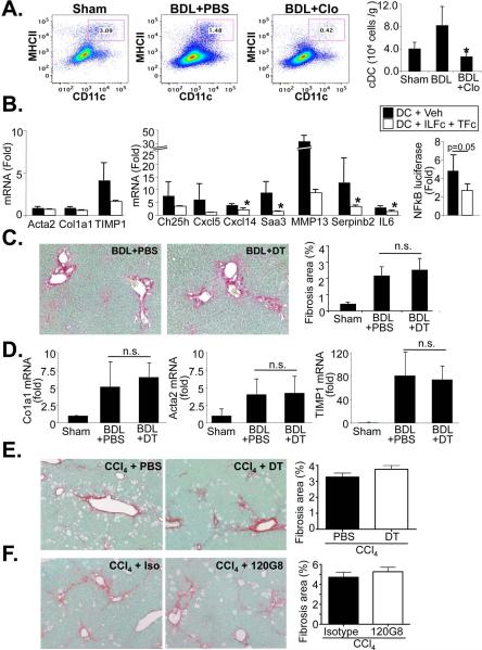
Figure 6. Dendritic cells moderately induce NF-κB dependent gene transcription in HSCs but do not contribute to BDL- and CCl4-induced liver fibrosis.
A. Bile duct ligated mice were injected every 5 days for a total of 3 injections with PBS (n=4) or clodronate (n=3) followed by quantification of cDC using CD11c and MHCII flow cytometry FACS plots show percentage of double-positive cells, the bar graph shows cell numbers per gram liver. B. mRNA expression of HSC activation markers (left panel), NF-κB responsive genes (middle panel) or NF-κB-driven luciferase activity (right panel) were determined by qPCR and NF-κB reporter assay, respectively, in HSC co-cultured with DC in the presence of antagonistic TNFRI-Fc and IL-1RI-Fc chimera (both 0.5 μg/ml) or vehicle (0.1%BSA in PBS) for 24h. C-D. Liver fibrosis was induced by BDL in CD11c-DTR bone marrow-chimeric mice. Chimeric male CD11c-DTR were treated with two injections of PBS (n=5) or diphtheria toxin (25 ng/g body weight) (n=6). Mice were sacrificed after 7 days. Deposition of fibrillar collagen was determined by Sirius Red staining (left upper panel) and morphometric quantification (right upper panel) (C). Expression of profibrogenic genes Acta2, Col1a1 and TIMP1 was determined by qPCR. Fold induction was calculated to sham-operated control (D). E. Chimeric CD11c-DTR mice (n=4 each group) were used for DC depletion (25 ng/g first two weeks, 10 ng/g last two weeks) and simultaneous liver fibrosis induction using CCl4 (0.5 μl/g, three time per week). Fibrosis was determined by Sirius Red staining. F. For pDC depletion C57Bl/6 mice (n=4) were injected every 48 hours with 120G8 antibody (500 μg/mouse) or isotype control during last two weeks of CCl4 induced liver fibrosis (0.5 μl/g, three time per week for four weeks). Fibrosis was determined by Sirius Red staining. * p<0.05

