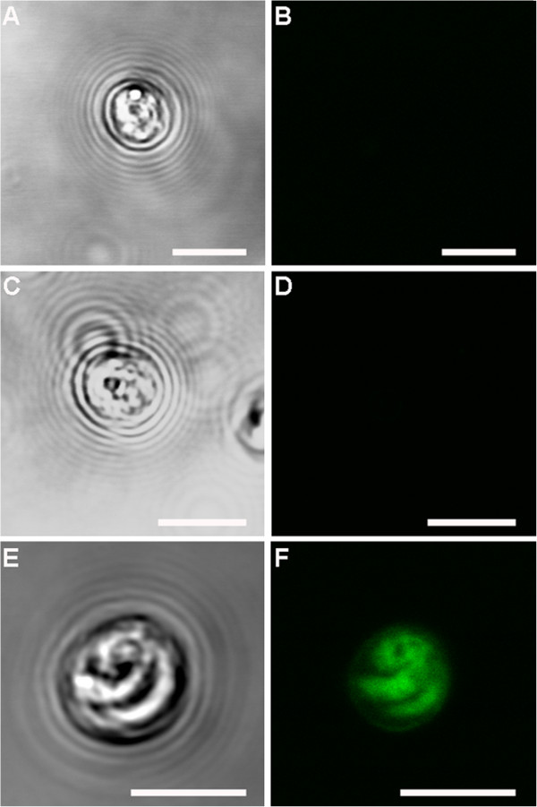
Figure 7.
Confocal observation of excysted oocysts control samples. A) excysted oocyst labelled with only primary antibody seen under transmitted light; B) corresponding confocal image, showing no immunolabeling. C) excysted oocyst labelled with only secondary antibody seen under transmitted light; D) corresponding confocal image, showing no immunolabeling. E) excysted oocyst labelled with both primary and secondary antibodies seen under transmitted light; F) corresponding confocal image, showing oocyst intensely immunolabelled. Scale bars: 5 μm.

