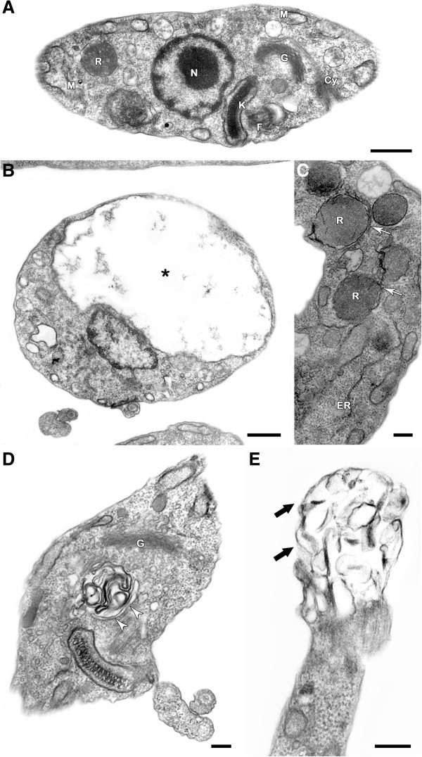
Figure 2.
Transmission electron microscopy analysis of T. cruzi epimastigotes treated with NQ1. (A) Untreated epimastigote showing normal ultrastructural aspect and presenting typical morphologies of the mitochondrion (M), kinetoplast (K), flagellum (F), nucleus (N), Golgi (G), reservosome (R) and cytostome (Cy). (B-E) The concentration of 0.3 μM NQ1 led to swelling in the mitochondrion (*), the formation of abnormal cytosolic membranous structures (white arrowheads) and the appearance of endoplasmic reticulum surrounding reservosomes (white arrows). Blebs (thick black arrows) was formed in the flagellar membrane of treated parasites. Bars = 500 nm (A, B, E) and 200 nm (C, D).

