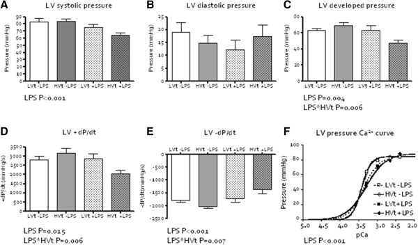
Figure 3.

Myocardial function ex vivo (LPS=lipopolysaccharide; LVt=low tidal volume (Vt) ventilation, HVt=high Vt ventilation). A. Left ventricular (LV) systolic pressure. B. LV diastolic pressure. C. LV developed pressure decreased by the interaction between LPS installation and HVt ventilation. D. LV contractility, measured as +dP/dtmax, decreased by the interaction between LPS installation and HVt ventilation. E. LV relaxation, measured as –dP/dtmin, decreased by the interaction between LPS installation and HVt ventilation. F. LV pCa50.
