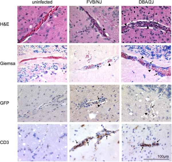
Figure 3.
Histological analysis of mouse brains. Brains from three infected male FVB/NJ and DBA/2J, and from control mice were prepared for histological analysis on day eight post-infection. Shown are representative images for sagittal brain sections that were stained with Giemsa or E&H for general brain pathology (first two rows). Parasites used in the infection express GFP and were visualized with an anti-GFP antibody (third row) and lymphocytes with an anti-CD3 antibody (last row). Arrowheads indicate infected red blood cells and arrows indicate leukocytes.

