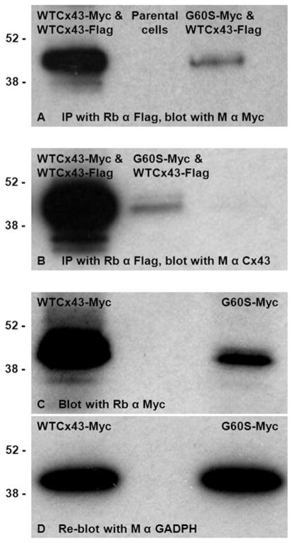
Fig. 4. G60S co-immunoprecipitates with WTCx43.
N2A cells were transiently co-transfected to express both WTCx43-Flag and WTCx43-Myc or WTCx43-Flag and G60S-Myc, and immunoprecipitated with a rabbit antiserum against Flag. The immunoprecipitants were immunoblotted with a mouse monoclonal antibody against Myc, confirming that WTCx43 and the mutant interact (A). Immunoblotting immunoprecipitates for Cx43 shows that the overall expression of Cx43 (WTCx43-Flag and either WTCx43-Myc or G60S-Myc) was lower for cells expressing G60S-Myc (B). Panels C and D are immunoblots of lysates of cells that were transiently transfected to express G60S-Myc or WTCx43-Myc, probed with a rabbit antiserum against Myc then reprobed with a monoclonal antibody against GAPDH. Note that the steady-state level of G60S-Myc is lower than that of the WTCx43-Myc (C), despite equal loading of the cell lysates as shown by blotting for GADPH (D). The positions of 38 and 52 kDa molecular markers are indicated.

