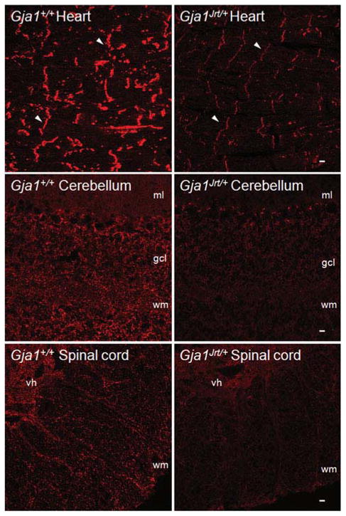
Fig. 5. Reduced Cx43-immunoreactivity in the heart, cerebellum and spinal cord of Gja1Jrt/+ mice.
These are confocal images of frozen sections from the heart, cerebellum and spinal cord of P40 Gja1Jrt/+ mice and their Gja1+/+ littermates. The sections were immunostained concurrently for Cx43 (red), and exposed for the same time to illustrate the diminished Cx43 staining in Gja1Jrt/+ tissues. In the heart, apposed membranes of cardiac myocytes from both Gja1+/+ and Gja1Jrt/+ mice are Cx43-positive. In the cerebellum, Cx43 is mostly localized in the granule cell layer (gcl) and white matter (wm) but not in the molecular layer (ml). In the spinal cord, Cx43 is more prominent in the gray matter of the ventral horn (vh) than in the white matter (wm). Scale bars: 10 μm.

