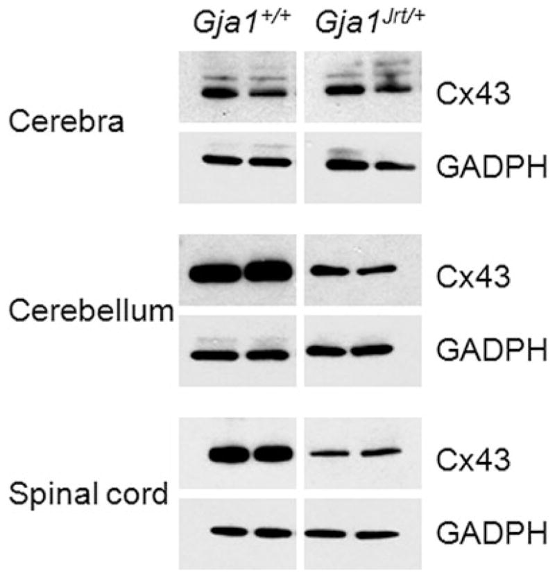
Fig. 6. Reduced Cx43 in the CNS of Gja1Jrt/+ mice.

These are immunoblots of individual cerebra, cerebella and spinal cords of P40 Gja1Jrt/+ mice and their Gja1+/+ littermates. Ten μg of protein/lane was separated by electrophoresis, transferred to a membrane, probed with a rabbit antiserum against Cx43, then re-probed with mouse monoclonal antibody against GADPH, to show that the loading was comparable. The level of Cx43 is lower in Gja1Jrt/+ cerebella (B) and spinal cords (C), but not cerebra (A). The larger bands of Cx43 in the cerebra are likely phosphorylated isoforms (Manias et al., 2008). The Gja1Jrt/+ and Gja1+/+ samples were on the same membrane, but the intervening lanes were spliced out.
