
Figure 1.
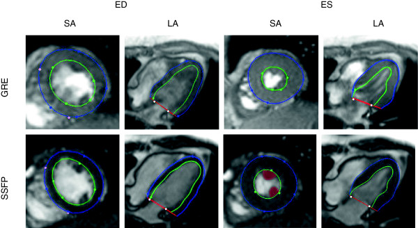
Image and shape differences for a volunteer imaged both with GRE (top), and SSFP (bottom), for the same short-axis (SA), long-axis (LA) planes at end diastole (ED) and end systole (ES). Green and blue contours and markers show the model’s endocardial and epicardial boundaries and guide points, respectively. Light color markers denote fiducial landmarks (right ventricular free wall insertion points, mitral valve hinge points) used to define the location of the model shape parameters in consistent positions relative to the anatomy of the heart. Papillary muscles are highlighted in red in the SSFP SA slice at ES.