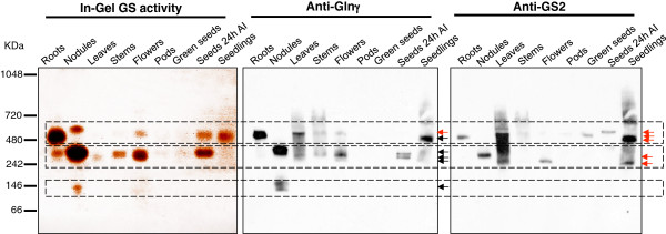
Figure 2.

Analysis of GS holoenzyme composition by Native polyacrylamide-gel electrophoresis in several organs of the plant. The organs analysed include roots, nodules, leaves, stems, flowers, pods, green seeds, seeds 24 hours after water imbibition (24 HAI) and seedlings. Equal amounts of protein (30 μg) were loaded in each lane. In-gel GS activity is revealed by the brown staining. The native gel was Western blotted using the anti-GS2 and anti-Glnγ antibody [27]. The same membrane was first incubated with the specific anti-GS2 antibody, stripped and re-probed with the Anti-Glnγ antibody. Three major regions of GS activity are indicated in the gel. Red and dark arrows indicate GS2 or GS1 protein complexes, respectively. The images are representative of at least three independent experiments.
