
Figure 1.
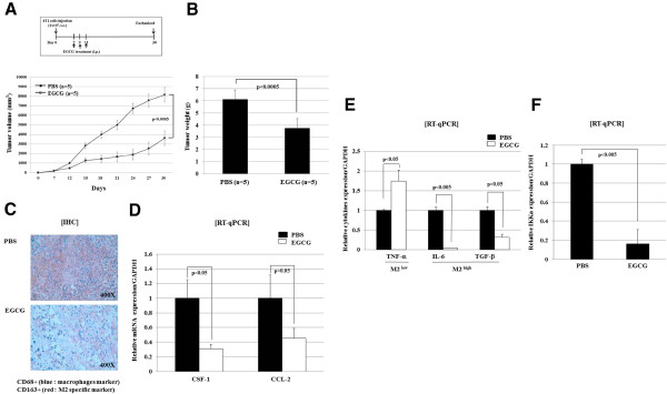
EGCG suppresses tumor growth, macrophages infiltration, and M2 polarization in in vivo murine breast cancer model. (A) Balb/c mice were challenged with 1× 10⁵ murine breast cancer cell lines, 4T1, by subcutaneous injection into the right flanks. EGCG (10 mg/kg) or PBS (as a negative control) was administrated intraperitoneally (i.p.) on days 7, 9, and 11. Tumour sizes were measured every week until day 30 post tumour challenge. (B) Mice were sacrificed and the tumors were dissected and the weight was measured. (C) For evaluation of tumor-associated macrophages and M2 macrophage infiltration, immunohistochemical staining for CD68 and CD163 was done using tumor tissue. (D) Tumor cells were isolated and total RNA was extracted and then subjected to RT-qPCR to measure the levels of CSF-1, CCL-2, and GAPDH. Histogram shows the relative expression of molecules compared to GAPDH as internal control. (E) Tumor-associated macrophages were isolated, and total RNA from these cells were extracted and subjected to RT-qPCR to detect levels of M2high cytokines (IL-6 and TGF-β) and M2low cytokine (TNF-α). Histogram shows the relative expression of molecules compared to GAPDH as internal control. (F) Tumor-associated macrophages were isolated and total RNA from cells were extracted and then subjected to RT-qPCR to detect the level of IKKα and GAPDH (internal control).