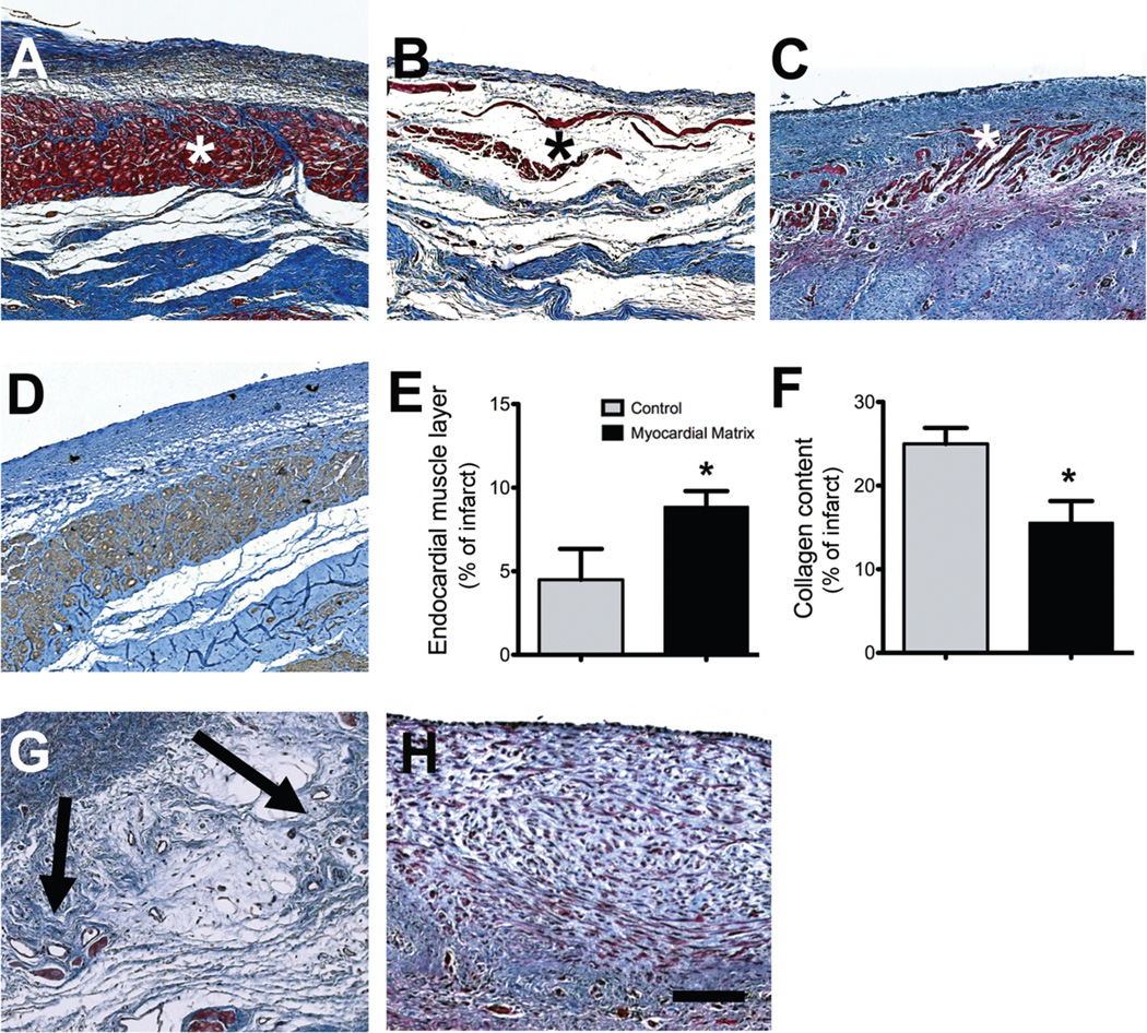
Fig. 3.
Histological characterization of infarcted pig hearts. (A to C) Masson’s trichrome staining images are representative of 6 matrix-injected pigs and 4 control animals. (A) Matrix-injected hearts had a distinct, thick endocardium (red, indicated with an asterisk). (B) Non-injected control animals had a loose fibrillar layer (blue) beneath the endothelium. (C) In saline-injected control animals, the endocardium was moderately thickened (red) (D) An adjacent tissue section for the matrix-injected animal in (A) stained for cardiac troponin-T, indicating the presence of cardiomyocytes. (E) Area of endocardial layer of muscle as a proportion of the infarct. (F) Percentage of collagen content in the infarcts. Data are means +/− SEM and were obtained from Masson’s trichrome slides (A to C). *p < 0.05, (Student’s t-test). (G and H) Matrix-injected hearts contained foci of neovascularization in the area below the endocardium (G, arrows) but none of the saline or non-injected control hearts showed these areas of neovascularization (H). Scale bar is 200 µm.

