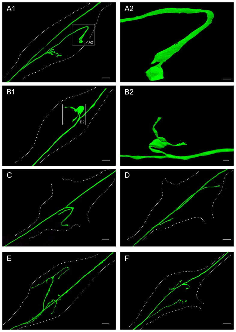
Figure 2. The AGR axon has a single (A-C), two (D-E) or three (F) projections from the main axon in the STG neuropil.
Dotted lines outline the neuropil area of the ganglion in all parts of the figure. A1. Volume-rendered view of LY dye-filled AGR with single projection, branching further into four sub-branches in the STG neuropil. The AGR soma is located in the stn outside the lower-left corner of the frame. Blend mode projection of 8 merged confocal image stacks, each consisting of 84 optical slices (acquired at resolution of 0.067µm x 0.067µm x 0.378µm). Scale bar is 30µm. A2 shows close-up of the boxed area in A1, showing the widened end of the AGR projection within the STG. Scale bar is 5µm. B1. Volume-rendered view of LY dye-filled AGR with single projection, branching further into two short sub-branches in the STG neuropil. The AGR soma is located in the stn outside the lower-left corner of the frame. Blend mode projection of 4 merged confocal image stacks, each consisting of 226 optical slices (acquired at resolution of 0.174µm x 0.174µm x 0.294µm). Scale bar is 30µm. B2 shows a volume-rendered surface projection of the boxed area in B1 from a different angle, emphasizing the big balloon-like widening (not the soma) of the axonal AGR projection. Scale bar is 10µm. C. Volume-rendered view of LY dye-filled AGR with single projection, branching further into two sub-branches in the STG neuropil. The AGR soma is located in the stn outside the lower-left corner of the frame. Blend mode projection of 5 merged confocal image stacks, each consisting of 169 optical slices (acquired at resolution of 0.068µm x 0.068µm x 0.462µm). Scale bar is 30µm. D. Volume-rendered view of LY dye-filled AGR with two simple projections in the STG neuropil. The AGR soma is located in the stn outside the lower left corner of the frame. Blend mode projection of 18 merged confocal image stacks, each consisting of 115 optical slices (acquired at resolution of 0.183µm x 0.183µm x 0.504µm). Scale bar is 30µm. E. Volume-rendered view of LY dye-filled AGR with two projections, each branching further into two or three sub-branches in the STG neuropil. The AGR soma is located in the stn outside the lower-left corner of the frame. Blend mode projection of 10 merged confocal image stacks, each consisting of 264 optical slices (acquired at resolution of 0.179µm x 0.179µm x 0.38µm), ventral view of same preparation as Figure 3. Scale bar is 30µm. F. Volume-rendered view of LY dye-filled AGR with three projections, one of them branching further into two sub-branches in the neuropil. The AGR soma is located in the stn outside the lower-left corner of the frame. Blend mode projection of 9 merged confocal image stacks, each consisting of 229 optical slices (acquired at resolution of 0.168µm x 0.168µm x 0.252µm). Scale bar is 50µm.

