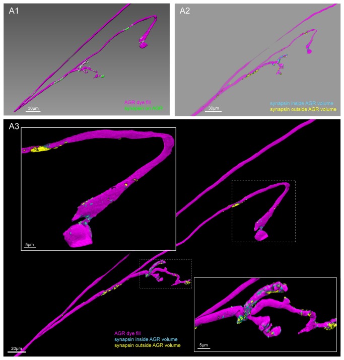
Figure 7. Clustered sites of putative chemical synapses are found in the AGR projections, but typically not in the AGR axon.
A1. Double labeling with an antibody against synapsin reveals patches of immuno-labeling on the LY dye-filled AGR projections. The image was processed to only show synapsin labeling in the AGR (see methods). Scale bar is 30µm. A2. Putative pre- and postsynaptic sites in the AGR projections in the same preparation. Different masking methods allow distinguishing between potentially presynaptic and postsynaptic sites in the AGR neuron (see methods). Synapsin labeling that mostly overlapped with the volume of the reconstructed AGR surface was classified as putative pre-synaptic (blue), and is found predominantly in the distal parts of the AGR process. Synapsin labeling that mostly overlapped with a thin shell around the AGR neuron was interpreted to be located in the processes of adjacent cells, marking putative post-synaptic sites in the AGR neuron (yellow). Scale bar is 30µm. A3. Overlay of the putative pre-and postsynaptic sites with the AGR projection (magenta). The close-up in the inserts reveals the distinct clustering in putative pre- and postsynaptic sites of the AGR projection and axon. Putative pre-synaptic sites in the AGR neuron are blue and putative post-synaptic sites are yellow. A1-A3 are blend mode projections of the same data set of 8 merged confocal image stacks, each consisting of 84 optical slices (acquired at resolution of 0.067µm x 0.067µm x 0.378µm).

