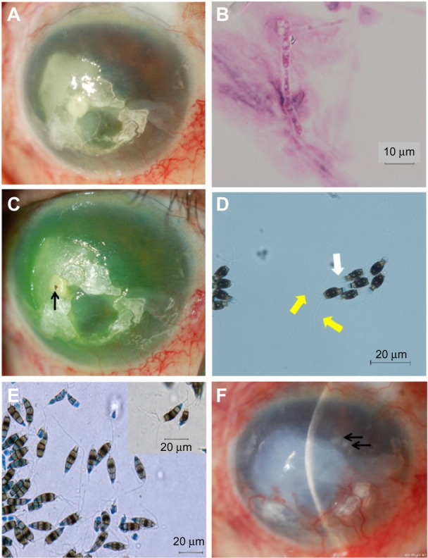
Figure 2.

Slit-lamp and light microscopy photographs.
Notes: (A) Oval infiltrate with irregular margins in the temporal half of the cornea at initial presentation. (B) Light microscopy of corneal scrapings taken from the right eye at initial presentation revealed uniformly thick septate hyphae. (C) There was a foreign body in the central infiltrate after corneal debridement (arrow). (D) Light microscopy that revealed conidia with three apical appendages (yellow arrows) and a single basal appendage (white arrow) (lactophenol cotton blue staining; ×400). (E) Microscopic findings of conidia produced on potato dextrose agar 1 month after incubation (lactophenol cotton blue staining; ×400). (F) Relapse of the fungal keratitis, 8 months after discharge (arrows).
