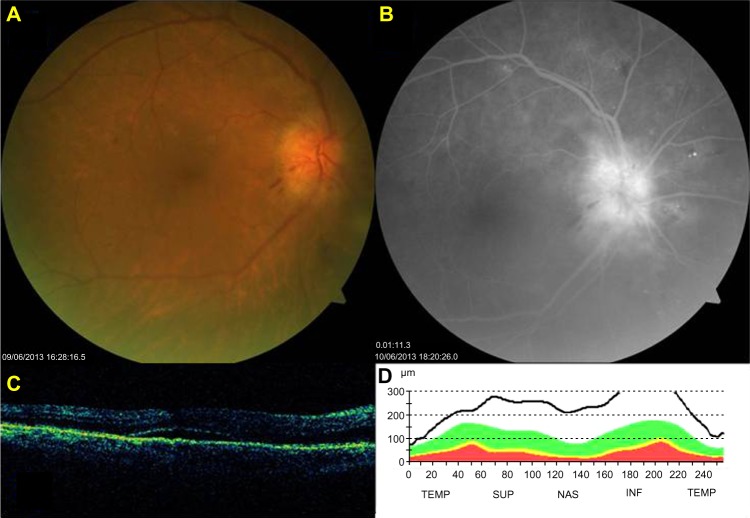
Figure 1.
(A–D) at presentation. (A) Fundus photography showing swollen optic disk. (B) Fluorescein angiography showing disk-vessel telangiectasia and disc leakage. (C and D) Optical coherence tomography scan showing a serous macular detachment. Marked edema of the optic disk and thickening of the retinal nerve-fiber layer.
Abbreviations: TEMP, temporal; SUP, superior; NAS, nasal; INF, inferior.

