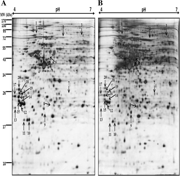
Figure 2.
Two-dimensional map of proteins from AGS gastric cancer cells treated with vitamin C. Proteins were isolated after exposure of the cells to A) control (only vehicle) and B) 300 μg/ml of vitamin C for 24 h and separated on IPG-strips with pH 4–7 in the first dimension, and then on 12% polyacrylamide gel on second dimension. The gels were silver stained.

