
Fig. 7.
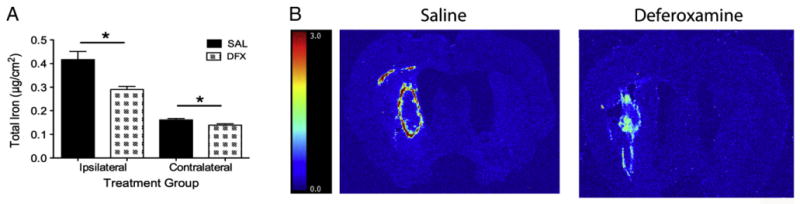
Total iron levels (mean±SEM) were quantified in the ipsilateral and contralateral hemispheres with RS-XRF. Giving DFX significantly lessened iron levels in both hemispheres (A; * p<0.05). Representative iron images are shown in B. The color scale is in μg/cm2.