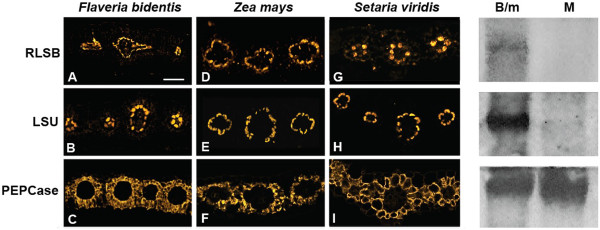
Figure 2.

Confocal Imaging showing co-localization of RLSB and Rubisco LSU in leaves of three C4 plant species. Column 1 (A, B, C): Flaveria bidentis, a C4 dicot. Column 2 (D, E, F): Maize (wild type line B73), a C4 monocot. Column 3 (G, H, I): Setaria viridis, a C4 monocot. Top row (A, D, G): leaf sections from the different C4 species were reacted with RLSB antisera. Middle row (B, E, H): leaf sections were reacted with Rubisco LSU antisera. Bottom row (C, F, I): leaf sections were reacted with PEPCase antisera (as an M-specific control). Note the centripetal chloroplast positioning within bundle sheath cells of F. bidentis, versus the centrifugal positioning in maize and S. viridis. Sections treated with indicated antisera were reacted with R-phycoerythrin (A and B) Alexafluor 584 (C to I) secondary antisera and captured using the 40X objective of a Zeiss 710 LSM Confocal microscope. Right Panels: Immunoblot of soluble B73 maize leaf protein extracts from mechanically separated cell populations produced using the leaf rolling method. B/m, cell population enriched in BS cells, with some M cells. Equal amounts of protein were loaded into each lane. M, purified M cells. Blots were incubated with antisera against RLSB, LSU, and PEPCase.
