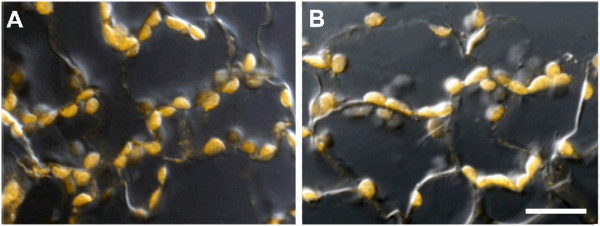
Figure 7.

Immunolocalization of RLSB and Rubisco LSU proteins in leaf sections of the C3 plant Arabidopsis. Panel A: Confocal/DICI image of Arabidopsis leaf section reacted with RLSB primary antiserum. Panel B: Confocal/DICI image of Arabidopsis leaf section reacted with LSU primary antiserum. Arabidopsis leaf sections were incubated with the indicated primary antiserum, and then Alexafluor 546 conjugated secondary antibody. Images were captured using the 40X objective of a LSM 710 “in tune” confocal microscope. Fluorescent immunolocalization was combined with bright field DICI to clearly show plastid localization for both proteins. bar = 20 μM.
