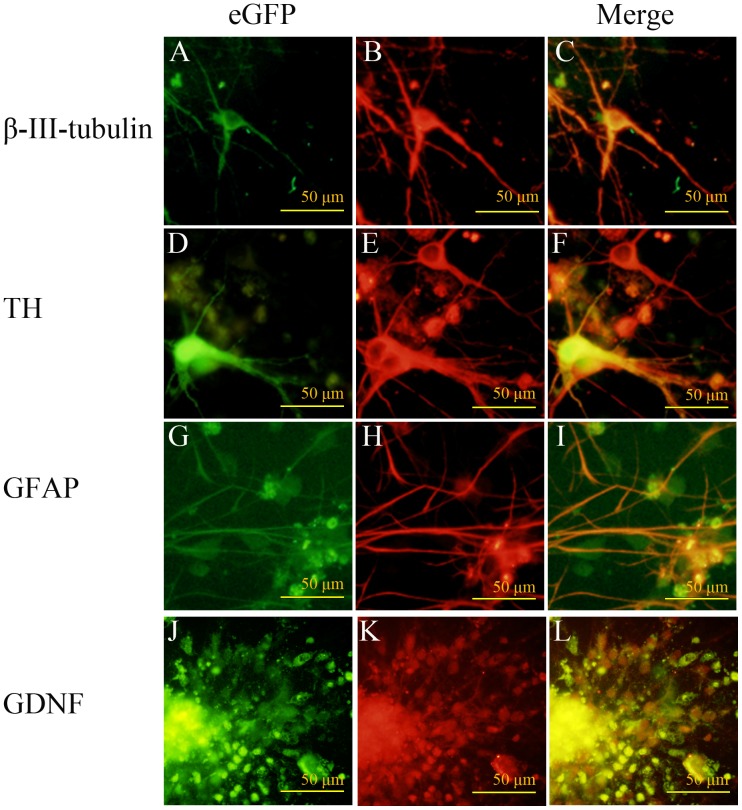
Figure 4. In vitro differentiation of SPIO, eGFP double-labeled GDNF-mNSCs.
Fluorescence photomicrographs showing SPIO, eGFP double-labeled GDNF-NSCs growing in a differentiation medium for seven days. eGFP-positive cells (A, D, G, J), immunoreactive for the neuronal markers β-III-tubulin (B) and TH (E), the astrocyte marker GFAP (H), and GDNF (K). Images are merged in the last column (C, F, I, L). Scale bar = 100 µm. Green: FITC, Red: TRITC.

