
Figure 1.
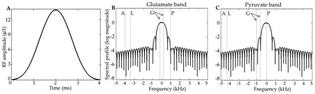
The spectrally selective hamming windowed-sinc RF pulse (Fig. A) and its spectral profile in log scale with passband placed on the glutamate band (Fig. B) and on the pyruvate band (Fig. C). The dashed lines marked G indicate the chemical shifts of [5-13C]glutamate and [1-13C]pyruvate in the passband with the resonances of acetylcarnitine, acetoacetate and citrate located between them. With the passband placed on the glutamate band, signal from [2-13C]pyruvate (line P) is suppressed. Lines L and A mark the locations of [2-13C]lactate and [2-13C]alanine, respectively. To image [2-13C]pyruvate or [2-13C]lactate, the passband is shifted to the corresponding frequency.