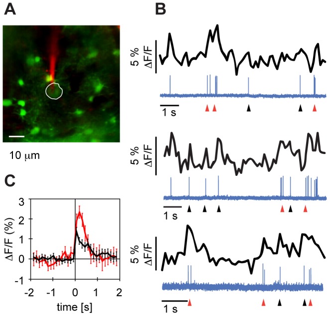
Figure 2. Action potentials are associated with fast calcium transients.

A Targeted juxtacellular recording of membrane potential from an unidentified HVC neuron (circumscribed in white) using a patch pipette containing 50 µM Alexa 594 (red). B. Three example traces of simultaneously recorded calcium signals (black) and juxtacellular membrane potential (blue, arbitrary units) of the neuron in A. Short increases in firing rate (low-frequency bursts, red arrows) but also single isolated spikes (black arrows) were associated with transient increases in fluorescence of up to 5%. C Event-triggered average (ETA) ΔF/F signals of the cell in A and B reveal average fluorescence transients of 1.4% for single spikes (black) and of 1.9% for low-frequency bursts (red, burst width = 200 ms) in the first bin after the event. The error bars indicate the standard error of the mean.
