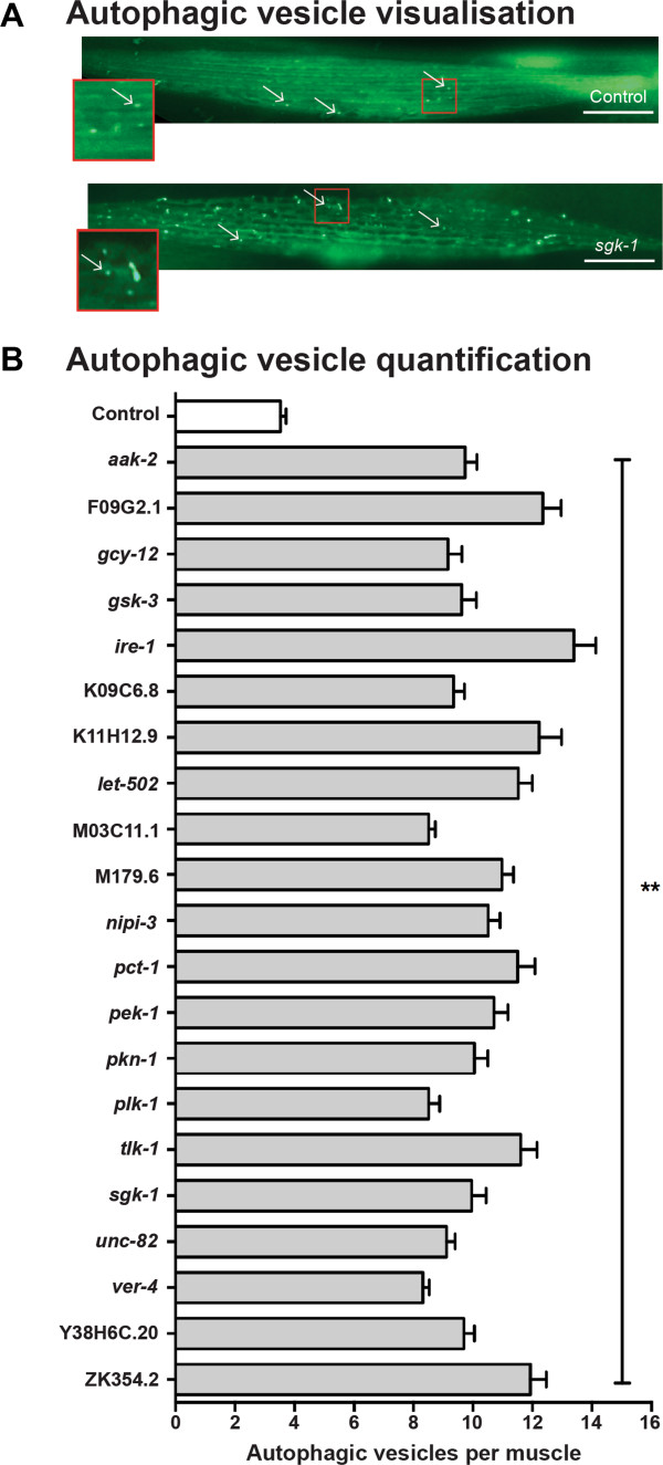
Figure 6.
Increased autophagic vesicles are present in muscles following knockdowns appear to trigger autophagy. (A) Sample images of GFP labelled autophagic vesicles showing normally low levels in a control RNAi treated KAG146 animal (left) and an experimental RNAi treated animal showing increased vesicles. RNAi treatment is indicated in the lower right corner. Scale bars represent 20 μm. (B) Each of the 21 kinase knockdowns that were identified as requiring autophagy to produce increased protein degradation displayed an increase in autophagic vesicles following 24 hours of acute RNAi treatment. Three independent experiments were performed (n = 20 each). Error bars represent standard error of measurement. ** p < 0.0001, one way ANOVA.

