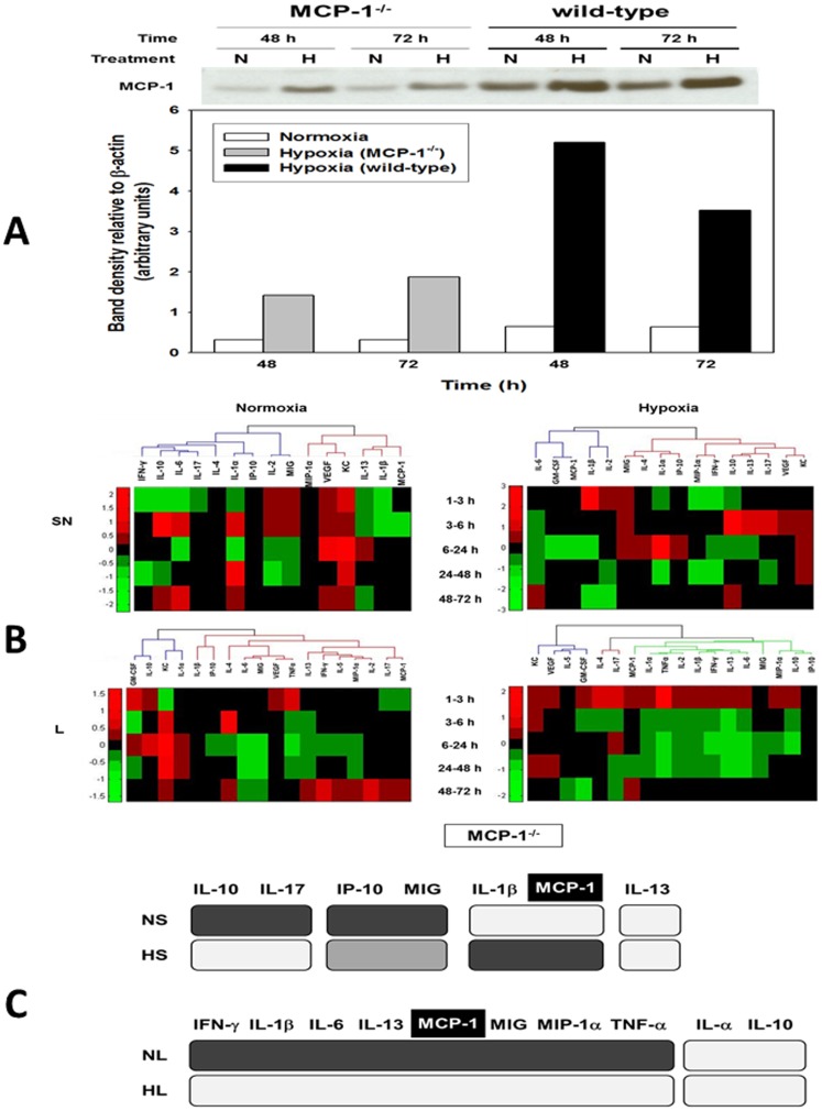
Figure 3. MCP-1 is a central component of the dynamic, multi-dimensional response of hepatocytes to cell stress.
Primary hepatocytes from wild-type and MCP-1−/− mice were cultured under normoxic (N) or hypoxic (H) conditions for 1–72 h, followed by Luminex™ analysis of 18 inflammatory mediators in both the supernatant (SN) and whole-cell lysate (L). The measurements were normalized and hierarchical and k-means clustering was performed as described in the Materials and Methods . (A) A representative Western blot showing MCP-1 protein expression in cell lysates from normoxic (N) and hypoxic (H) MCP-1−/− and wild-type mouse hepatocytes (48 and 72 h) and densitometric analysis as described in the Materials and Methods . (B) Hierarchical clustering over fold changes in MCP-1−/− hepatocytes (normoxia vs. hypoxia): Fold change values for each inflammatory mediator ranging from large negative (green) to large positive values (red) are shown. No fold changes (zero values) are represented in black. (C) Comparison between meta-clustering analysis outcomes in normoxia and hypoxia (see Materials and Methods ). The shading of the boxes indicates the grouping of mediators that exhibited the same segregation pattern across all methods. For each experimental condition (NL, HL, NS and HS), only the mediators appearing in each consensus are shown. For comparison between experimental conditions only mediators common to both consensuses are shown.

