
Fig. 3.
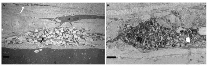
Transmission electron microscopic findings. (A) Keratocyte distended by membrane-bound intracytoplasmic vacuoles containing electron dense fibrillogranular material (black arrow). Vacuole containing dense fibrillogranular material in the interstromal lamellae (white arrow). Asterisk indicates relatively normal keratocyte (scale bar, 2 µm). (B) Keratocyte with vacuoles containing dense osmophilic whorls (scale bar, 0.5 µm).