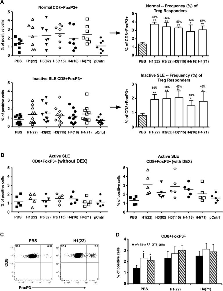
Figure 2.
Durable induction of CD8+FoxP3+ cells in vitro by low-dose histone peptides. (A) Fresh PBMCs samples derived from healthy subjects and inactive SLE patients were cultured as described in Figure 1A and stained for CD8 and FoxP3. Y-axes show % of FoxP3 positive cells among viable T cells gated for being CD8+. pCntrl = control peptide A; horizontal lines indicate the mean. Similar to right panels in Figure 1A and as explained in the corresponding legend to Fig. 1A, the right panels here (next to →) show the degree of CD8+FoxP3+ responses (bars = mean ± SD), except for the bar for PBS, and the frequency (% numbers above bars) of positive CD8 Treg responders for each peptide. ** p < 0.01, * p < 0.05. n= 5 to 10. (B) Fresh PBMCs from active SLE patients were cultured with histone peptide epitopes and IL2, in the presence or absence of Dexamethasone (DEX) for 7 days, and then stained for CD8 and FoxP3. (C) Representative Dot Plots show induction of CD8+FoxP3+ T cells in PBMC after culturing with the histone peptide epitope H122-42 for 7 days. (D) Shows the effects of TGF-β (Tβ) or Retinoic Acid (RA) on induction of CD8+FoxP3+ Treg cells by two of the peptide epitopes, n = 3, * p < 0.05.

