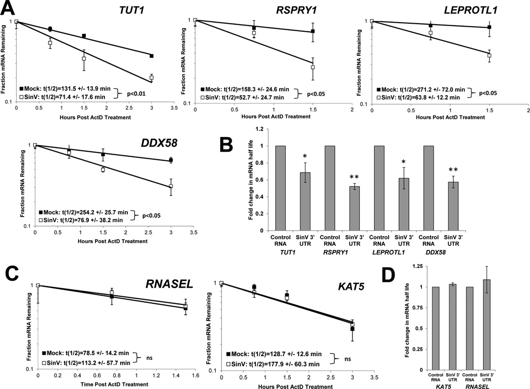
Figure 2. SinV infection influences the stability of some but not all cellular mRNAs.
At 24 hpi with SinV, 293T cells were treated with actinomycin D and the relative levels of the indicated mRNAs were assessed at the designated time points following shut off of transcription using qRT-PCR to determine mRNA half-lives. Panel A depicts mRNAs that were destabilized during SinV infection while panel C contains mRNAs whose stability was not affected. Representative decay curves are shown with standard deviation of experimental measurements and the average half-lives are reported with standard deviations from three independent experiments. In Panels B and D, 293T cells were transfected with equimolar amounts of either a control RNA (GemA60) or the SinV 3’UTR RNA. At 4.5 hrs post transfection, cells were treated with actinomycin D and the relative levels of the indicated mRNAs were assessed by qRT-PCR. Average fold change in half-lives are reported with standard deviations from two independent experiments. * p<0.05; ** p<0.01.

