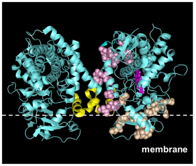
Figure 4. Functional networks of CYP2C9.

Dimerization and membrane-binding networks were mapped on the crystal structure model of a CYP2C9 dimer (PDB code 1R9O). The dimerization network is shown in pink, the membrane-binding network is shown in orange, and the heme group is shown in magenta on the right monomer. The F-G loop is shown in yellow. The position of the membrane line was qualitatively positioned based on the location of the residues in the membrane-binding networks.
