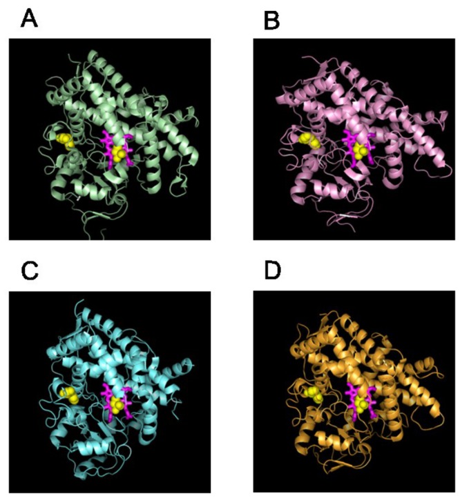
Figure 5. Co-evolved residues in the heme-binding networks.

Residues identified as belonging to heme-binding networks were mapped to the molecular structures of CYP1A (PDB code 2HI4; A), CYP2C (PDB code 1R9O; B), CYP3A (PDB code 1W0E; C), and CYP2D (PDB code 2F9Q; D). The heme molecule is shown in stick representation, and residues belonging to the functional networks are shown as yellow spheres. Note that this network consists of both co-evolved residues that are in direct proximity to the heme as well as residues that are located distal to the heme.
