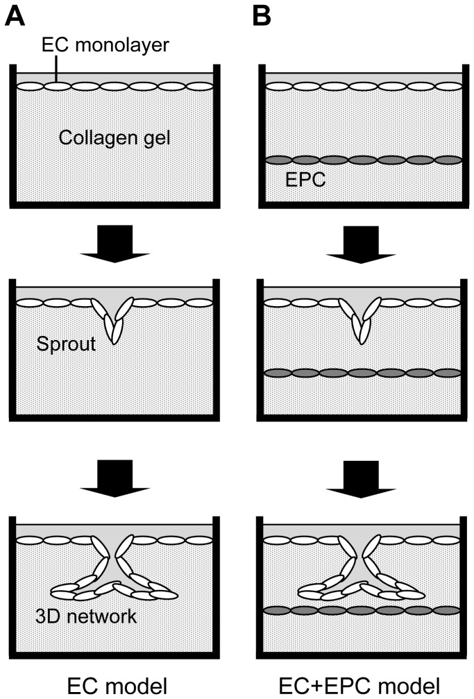
Figure 1. Three-dimensional endothelial network models.
(A) In the EC model, ECs were seeded onto collagen gel. The EC monoculture served as a control. (B) In the EC+EPC model, EPCs were sandwiched with double layers of collagen gel. ECs were then cultured on the top of the upper collagen gel layer. In each model, some ECs in a confluent monolayer invaded the underlying collagen gel with the addition of bFGF (Sprout) and formed 3D endothelial networks in culture (3D network).

