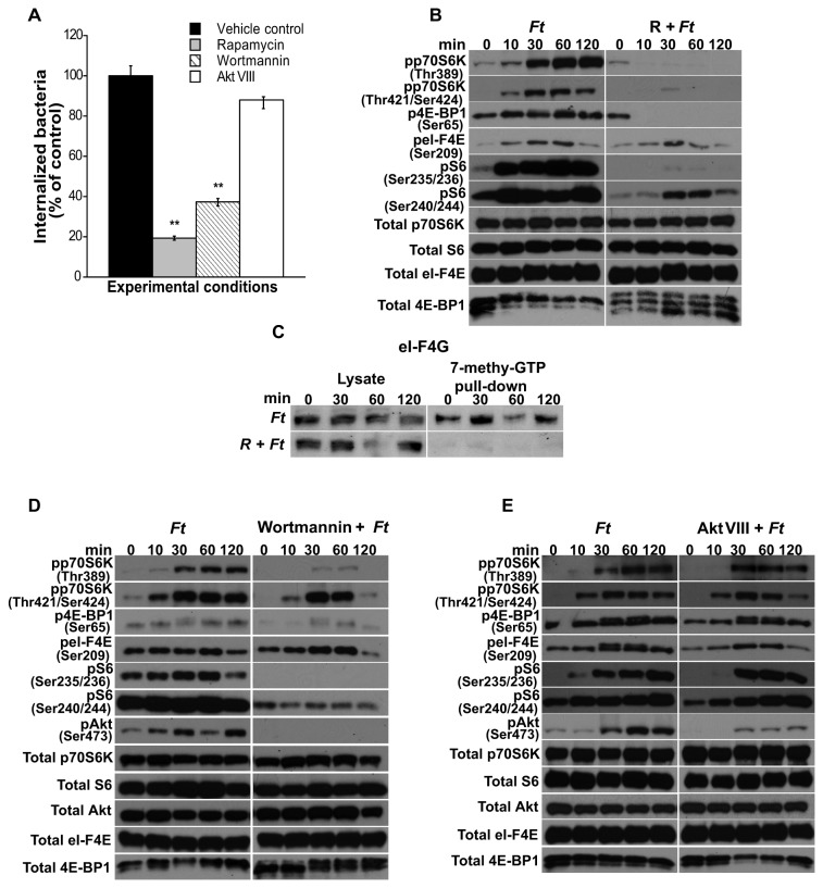
Figure 1. Effect of mTOR, PI3K and Akt inhibitors on the internalization of F. tularensis LVS.
(A) Peritoneal macrophages derived from WT mice were pretreated or not with rapamycin (50 µg/ml; 3 h), wortmannin (100 nM; 1 h) or Akt VIII (500 nM; 1 h) and infected with freshly harvested F. tularensis LVS (MOI=20) for 90 min to assess bacterial invasion. Values are the mean ± SEM of 5 independent experiments, each done in triplicate; **p < 0.001; *p < 0.05 compared with infected control cells treated with DMSO. Peritoneal macrophages derived from WT mice were pretreated or not with the inhibitors as described above, exposed to F. tularensis LVS for 0-120 min and then lysed. (B, D, E) Total p70S6K, S6, 4E-BP1, eI-F4E and Akt, and phosphorylated p70S6K (Thr389 and Thr421/Ser424), 4E-BP1 (Ser65), S6 (Ser235/236 and Ser240/244), eI-F4E (Ser209) and Akt (Ser473) were assessed by Western analysis. Samples analyzed contained an equal amount of protein. Unstimulated control cells (time 0) were incubated with the respective inhibitors for the correspondent pre-incubation period. Prior to the addition of bacteria, cells were not washed including unstimulated controls. Unstimulated cells served as negative controls. (C) Peritoneal macrophages were pretreated with rapamycin (50 µg/ml) and exposed to F. tularensis LVS for 0-120 min. An equal amount of protein from each lysate was pulled-down using 7-methyl GTP sepharose beads. Pull-down products were assessed for eI-F4G by Western analysis. All gels are representative of three to five independent experiments.

