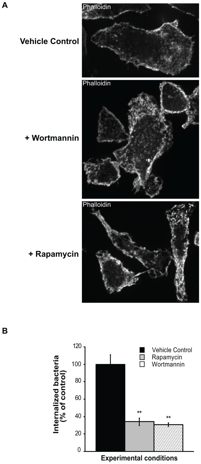
Figure 2. Effects of wortmannin and rapamycin on actin cytoskeleton and F. tularensis entry into cells.

(A) Peritoneal macrophages were treated with vehicle control, wortmannin (1 h) or rapamycin (3 h) and stained with fluorescent β-phalloidin to detect F-actin. Wortmannin and rapamycin significantly alter the architecture of the actin cytoskeleton and induce numerous thick, short filaments and small patches distributed throughout the cell. Representative cells are shown. (B) Isolated macrophages were treated with vehicle control, wortmannin (for 1 h) or rapamycin (for 3 h), and then F. tularensis was added for 90 min. Cells were processed by immunofluorescence with an F. tularensis anti-LPS antibody to detect internalized bacteria. One hundred (100) cells for each treatment were counted in randomly selected fields. The number of bacteris in treated cells is expressed as percent normalized to the control (100%). The p values of rapamycin- and wortmannin-treated cells are <0.001 relative to control.
