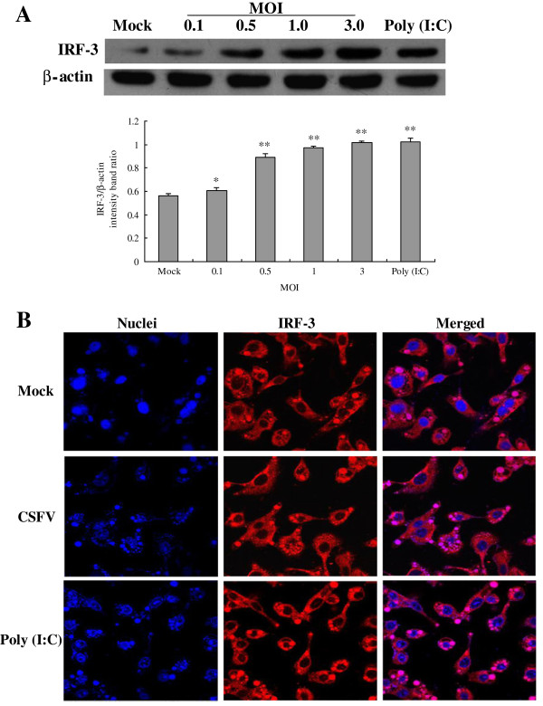
Figure 3.
Expression and nuclear translocation of IRF-3 after CSFV infection in porcine alveolar macrophages. (A) Expression of IRF-3 was measured by Western Blotting with antibodies specific for IRF-3, and the cells were treated as demonstrated in Figure 1. (B) Indirect immunofluorescence analysis was used to measure cellular localization of IRF-3 in CSFV-infected PAMs. Cells were mock treated, poly (I:C) stimulated, or infected with Shimen isolates at an MOI of 3. At 24 hpi, cells were fixed and the localization of IRF-3 (red) was observed by fluorescence microscope using immunofluorescence stain with anti-IRF-3 and QDs-SA 605-conjugated and biotinylated secondary antibodies. Nuclei were stained with DAPI. Bar, 10 μm. The experiment was repeated three times and the figure shows a representative experiment. An asterisk indicates a statistically significant difference from uninfected cells, * P < 0.05 and ** P < 0.01.

